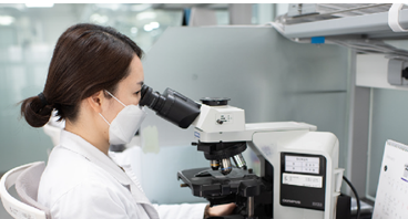
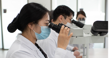
Pathologic Laboratory
Pathology Test
Pathology accurately classifies and diagnoses diseases by precisely deciphering the physical, immunological, and molecular biological changes in the body due to diseases.
-
01. Standardized Sample Reception System
Standardized sample reception system at 16 branches nationwide -
02. Quick Diagnosis System
Established a quick diagnosis system with pathological diagnosis coding -
03. Advisory System
Advisory system linked with university hospitals
Accurate and prompt results are provided by establishing a pathological analysis system with skilled and experienced specialists and advanced equipment
- 01
Optimized Facilities for Tests
- Equipped with latest equipment
- Analysis equipment of the best facilities
- Accurate results by outstanding talents
- 02
Prompt and Accurate Diagnosis
- Standardized reception system
- Pathological diagnosis coding
- Annual advisory system of branches
- Advisory system linked to university hospitals
- 03
Endless Research and Quality Control
- Participation in conferences in Korea and overseas and research council activities
- Implemented Daily Quality Control Conference
- Certified by the Korean Society of Pathologist (KSP)
- Certified for Quality Management System (ISO)
Major Cytopathology Tests
- - Exfoliative cytology: Observation of cells that naturally falls off such as vaginal discharge, sputum, and urine
- - Fine-needle aspiration biopsy: Poking the lesion area with a needle and collecting cells for analysis
- - Liquid-based cytology: A test increasing the diagnosis rate and reducing the loss of cells through the special preservation solution
Major Histopathology Tests
- Pathological diagnosis of tissues obtained through excision or biopsy
-
Thinprep2000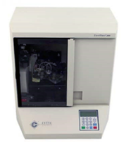
Cytopathology
Cytopathology includes the fine-needle aspiration biopsy that observes cervical cells or cells from sputum, urine, and body fluid as well as aspirated cells for the thyroid, breasts, and lymph nodes by poking the lesion with a fine needle. Liquid-based cytology involves shaking the collection tool in the vial containing the cell preservation solution to wash the cells and then processing the preservation solution using the dedicated equipment to form a uniform and thin cell population smeared with a diameter of 2 cm. This has low loss of cells, prevents degeneration, and increases the accuracy of the diagnosis.
-
Tissue Tek Prisma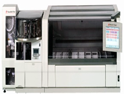
Cytopathology
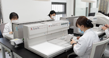
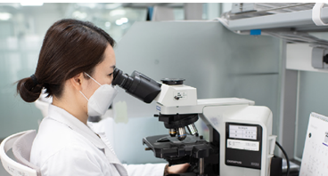
The slides are placed in a rack for H&E stain or pap stain, and the automatic glass cover slipper next to it attaches the glass cover to the section or smeared surface. The glass cover protects the section or smeared surface and prepare microscope slides. The prepared slides are examined by pathologists with an optical microscope.
-
Tissue Tek VIP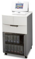
Pathology Test
This equipment performs pre-processing of the fixed tissue through dehydration (alcohol), transparency (xylene), and penetration (paraffin) processed in 12 hours. Tissue slides prepared through the pre-processing are appropriate for microscopic tests.
-
Tissue Tek TEC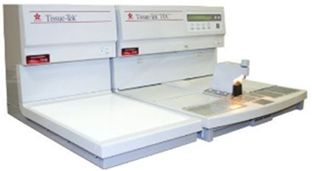
Pathology Test
This equipment prepares pre-processed tissues into blocks that can easily be sliced using paraffin.
-
Leica RM2255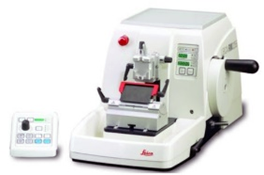
Pathology Test
This equipment prepares the internal structure of the tissue to be observed accurately under a microscope by slicing the prepared blocks into 4 μ thickness.
-
Leica IP C Inkjet printer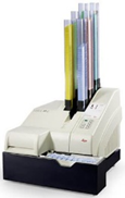
Pathology Test
This equipment automatically labels cassettes with pathology numbers assigned to each sample.
-
PPM-SX slide printer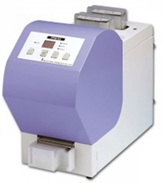
Pathology Test
This equipment automatically labels slides with pathology numbers and names assigned to each sample.
